Solid Tumors and Metastasis

Solid Tumors, Metastasis & Therapy Resistance

Solid Tumors and Metastasis
Hematopoietic Stem Cells and Leukemia

Hematopoietic Stem Cells and Leukemia

Hematopoietic Stem Cells and Leukemia
Stem Cells and Tumor Microenvironment

Stem Cells and Tumor Microenvironment

Stem Cells and Tumor Microenvironment
HI-STEM Group Leaders

HI-STEM Group Leaders

HI-STEM Group Leaders
PlayPause
previous arrow
next arrow

Pluripotent cell lines can be differentiated into various blood cell types via the process of embryoid body formation. The burst-forming erythroid unit (BFU-e) comprises a type of red cell progenitor while the colony forming unit granulocyte-macrophage (CFU-GM) is a bi-potent myeloid progenitor cell capable of making both granulocytes and macrophages.

During embryonic development, HSCs are generated from pluripotent cells in a series of progressive differentiation steps. This process can be modeled in vitro by directing the differentiation of pluripotent cell lines, such as embryonic stem (ES) cells or induced pluripotent stem cells (iPSCs), towards the formation of the hematopoietic lineages. The in vitro specification of hematopoietic lineages from ES cells and patient-specific iPSCs closely mimics the process via which HSCs are generated during development, and holds enormous potential for disease modeling and for the eventual scalable production of differentiated cells for regenerative therapy. However, a number of differences exist between the normal developmental process and the in vitro differentiation procedure. The most notable of these differences is the current inability to generate in vitro, HSCs that have the same functional properties as their in vivo specified counterparts. One of the current focuses of the Experimental Hematology group is to better understand the important molecular and biological differences between hematopoietic specification in vitro and in vivo, and to attempt to apply this knowledge towards developing better methodologies for the generation of functional HSCs from ES cells and iPSCs.


Key Publications on pluripotent cell lines and HSC specification

1. §Müller L.U.W., §Milsom M.D., Harris C.E., Vyas R., Brumme K.M., Parmar K., Moreau L.A., Schambach A., Park I.H., London W.B., Strait K., D’Andrea A., Daley G.Q. and Williams D.A. (2012). Overcoming reprogramming resistance of Fanconi anemia cells. Blood. 119 (23): pp5449-57. §Equal contribution.

2. Ghiaur G., Ferkowicz M.J., Milsom M.D., Bailey J., Cancelas J.A., Yoder M.C. and Williams D.A. (2008). Rac1 is essential for intraembryonic hematopoiesis and for the initial seeding of the fetal liver with hematopoietic progenitor cells. Blood, 111 (7): pp3313-21.


With advancing age, most regenerating organs suffer a cumulative decline in functional capacity that is associated with an increase in age-associated diseases such as cancer. These phenomena are thought to be driven by defects at the level of adult stem cells, whose progressive attrition with age is likely to precipitate tissue dysfunction. Adult stem cells are also the likely cell of origin for many cancers. There are multiple lines of evidence implicating the accumulation of DNA damage within adult stem cells as causative for these age-associated phenotypes. Firstly, many accelerated ageing disorders (progeroid syndromes), such as Hutchinson-Gilford progeria, Werner syndrome, Bloom’s syndrome, xeroderma pigmentosum, Cockayne syndrome, dyskeratosis congenita and Fanconi anemia, are caused by defects leading to elevated levels of DNA damage and/or in the compromised repair of DNA damage. Secondly, DNA damage has been shown to provoke cell fate decisions in adult stem cells that would be consistent with depletion with age. These include apoptosis, senescence, differentiation and malignant transformation. Thirdly, DNA mutations have been shown to accumulate in adult stem cells in an age dependent manner, with some estimates predicting that human HSCs acquire around 14 mutations per year per stem cell, which equates to an average mutation burden of around 700 mutant alleles in each HSC by the time an individual reaches the age of 50. Finally, pre-malignant oncogenic mutations have been found to exist in the normal stem cell pool of aged individuals. These mutations impact on HSC behavior, and can drive the non-malignant clonal outgrowth of individual HSC clones, which is associated with an increased risk of developing hematologic malignancy.

Although DNA damage within stem cells is well accepted as an important mechanistic cause of tissue ageing, the main physiologic source of DNA damage within the stem cell compartment is not clear. A more thorough understanding of the environmental factors that are responsible for DNA damage in these rare but important cell populations would potentially allow better assessment of the risk of a individual developing either cancer or other age related pathologies. One could also imagine that this knowledge would also facilitate the development of therapeutic strategies to prevent or delay this process.

Environmental stress is a biologically relevant mediator of DNA damage in the HSC compartment

A rude awakening

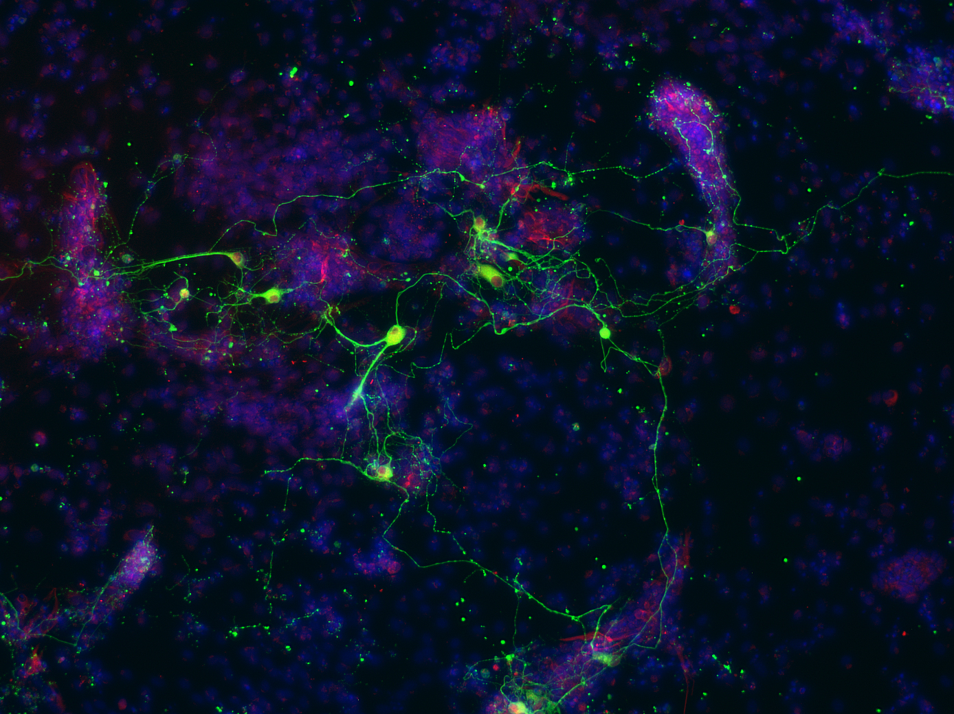
A rude awakening for stem cells: Various forms of environmental stress, such as infection, inflammation or blood loss, can force HSCs to leave their so-called “dormant” state and enter into active cell cycle in order to replenish mature blood cells. This period of forced exit from dormancy corresponds to conditions of high replication stress within HSCs, leading to DNA damage and HSC depletion.

A phenomenon known as replication stress is one potential source of DNA damage that is broadly applicable to most cell types across all individuals. Replication stress can be defined as any setting where the DNA replication fork stalls, increasing the likelihood of DNA strand breaks if the fork subsequently collapses. A number of biological phenomena can contribute to elevated levels of replication stress, including nucleotide pool imbalances, the presence of DNA adducts, hybrid DNA-RNA structures called R-loops and the presence of highly repetitive DNA sequences or complex secondary structure, all of which can inhibit the progression of the replication fork along DNA during S phase. However, it goes without saying that replication stress is only relevant in the context of actively proliferating cells and this poses a problem when we come to consider HSCs, which are characterized by very low levels of cell cycle activity under homeostatic conditions. In fact, the functional potency of HSCs has been shown to positively correlate with this status of long-term quiescence, also known as dormancy. We therefore hypothesized that HSC dormancy might be a route via which the genomic and associated functional integrity of HSCs is preserved during ageing, by acting to limit replication stress. This dormant state can however be broken under certain physiologic conditions, such as during infection or following blood loss, where HSCs are forced into cycle in order to regenerate the blood system. In a recent publication in the journal Nature1, we have shown that the exposure of mice to various forms of environmental stress can lead to DNA damage in HSCs as a result of their exit from their homeostatic dormant status. Importantly, the biological relevance of this finding to ageing was made clear when we observed that chronic stress led to a depletion in the number of functional HSCs, resulting in a phenotype akin to accelerated ageing of the blood system.

 The Fanconi anemia DNA repair pathway is an important route via which stress-induced DNA damage is resolved.

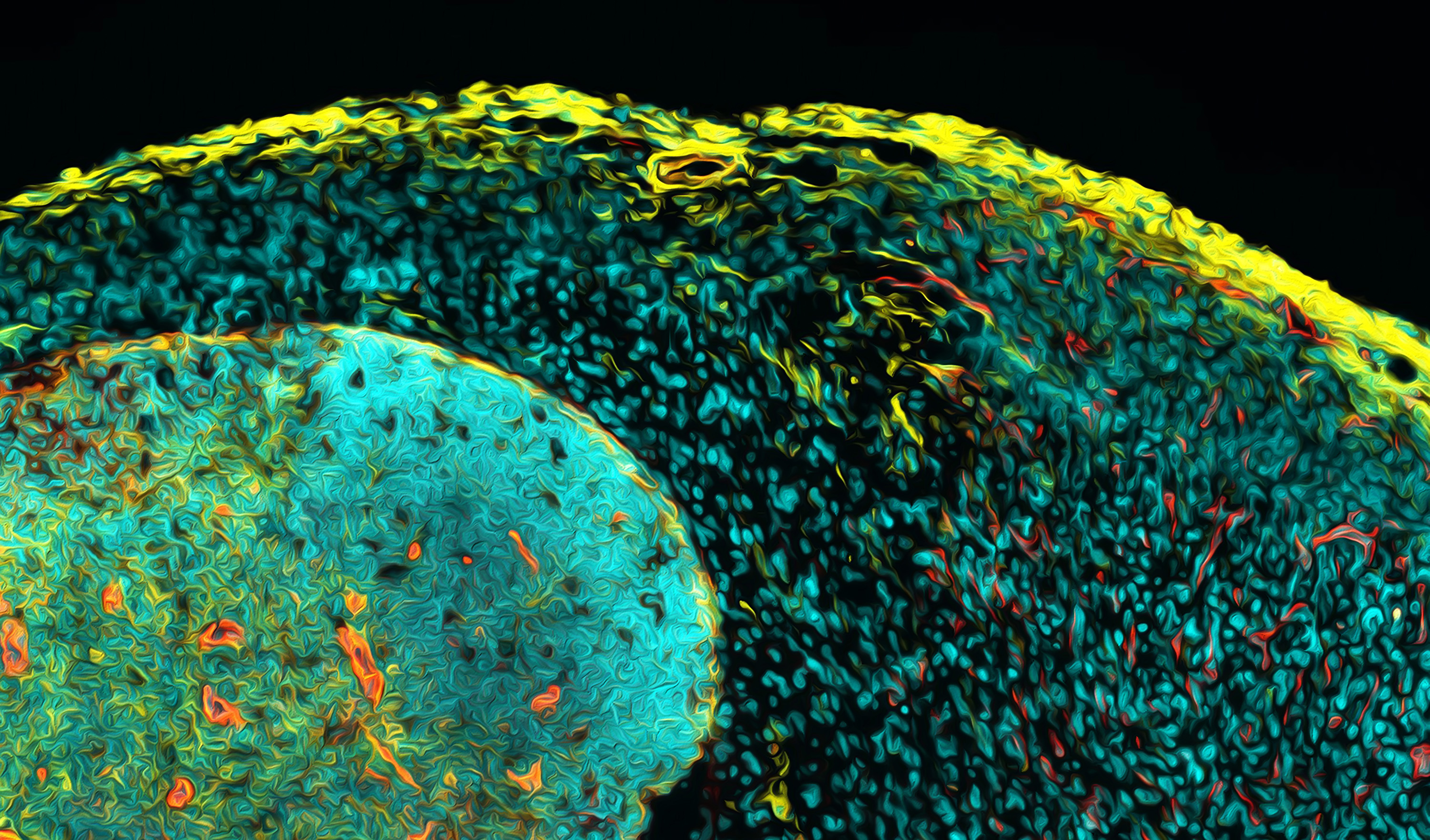
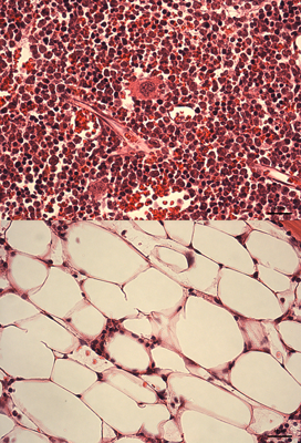
Stress hematopoiesis leads to bone marrow failure in DNA-repair defective Fanconi anemia knockout mice: Fanconi anemia knockout mice subject to chronic inflammatory stress (bottom panel) demonstrate severe bone marrow aplasia compared to their wild type counterparts subject to the same treatmenregimen (top panel). The image shows H&E stained bone marrow sections, with different types of blood cells being clearly very abundant in the wild type marrow, while in the FA knockout marrow, the blood cells have been depleted to be replaced by adipocytes.

Fanconi anemia (FA) is the most common inherited bone marrow failure syndrome, resulting from inactivating mutations in any of up to 18 genes, which together form part of an epistatic signaling pathway. This pathway is involved in resolving certain forms of DNA damage related to stalled replication forks. Most FA patients develop early onset bone marrow failure that is thought to be driven by accelerated depletion of the HSC pool, leading to the disease being considered as a type of segmental progeria syndrome (reviewed in 2). In line with this accelerated ageing phenotype, FA patients also have an extremely high risk of developing leukemias and solid tumors. Mouse knockout models of FA have exactly the same DNA repair defect as that observed in human patients, but they never develop bone marrow failure, even at extreme old age3. This is likely due to the FA mice not encountering the source of DNA damage that is responsible for HSC attrition in FA patients during ageing. We hypothesized that HSC dormancy may protect FA knockout murine HSCs from DNA damage and that environmental stress may ultimately be the missing source of DNA damage in experimental mice. In line with this concept, we could show that HSCs from FA knockout mice demonstrated elevated levels of DNA damage in vivo in response to various forms of physiologic stress, suggesting that the FA pathway is an important route via which this damage is resolved. Importantly, if we repeated the chronic stress treatment that resulted in accelerated ageing within the hematopoietic system of wild type mice, this led to an almost complete exhaustion of the HSC pool in FA mice and precipitated a highly penetrant bone marrow failure phenotype, highly reminiscent of that observed in patients1. We believe that this new model will not only allow us to better understand the etiology of this devastating disease, but will also permit us to more broadly gain new insight into the process of ageing and malignant transformation across the general population.

Key Publications on DNA damage in HSCs and Fanconi anemia

1. Walter D., Lier A, Geiselhart A., Thalheimer F.B., Huntscha S., Sobotta M.C., Moehrle B., Brocks D., Bayindir I., Kaschutnig P., Müdder K., Klein C., Jauch A., Schroeder T., Geiger H., Dick T.P., Holland-Letz T., Schmezer P., Lane S.W., Rieger M.A., Essers M.A.G., Williams D.A.W., Trumpp A.and Milsom M.D. (2015). Exit from dormancy provokes DNA damage-induced attrition in haematopoietic stem cells. Nature, 520(7548): pp549-552).

2. Geiselhart A., Walter D., Lier A. and Milsom M.D. (2012). Disrupted signalling through the Fanconi anemia pathway leads to dysfunctional hematopoietic stem cell biology: underlying mechanisms and potential therapeutic strategies. Anemia, 2012: 265790.

3. Kaschutnig P., Bogeska R., Walter D., Lier A., Huntscha S. and Milsom M.D. (2015). The Fanconi anemia pathway is required for efficient repair of stress-induced DNA damage in hematopoietic stem cells. Cell Cycle, 14(17): pp2734-2742.

4. §Müller L.U.W., §Milsom M.D., Harris C.E., Vyas R., Brumme K.M., Parmar K., Moreau L.A., Schambach A., Park I.H., London W.B., Strait K., D’Andrea A., Daley G.Q. and Williams D.A. (2012). Overcoming reprogramming resistance of Fanconi anemia cells. Blood. 119 (23): pp5449-57. §Equal contribution.

5. Milsom M.D., Schiedlmeier B., Bailey J., Kim M.O., Li D., Jansen M., Ali M.A., Kirby M., Baum C., Fairbairn L.J. and Williams D.A. (2009). Ectopic HOXB4 overcomes the inhibitory effects of tumor necrosis factor-α on Fanconi anemia hematopoietic stem and progenitor cells. Blood, 113 (21): pp5111-20.

6. Milsom M.D., Lee A.W., Zheng Y. and Cancelas J.A. (2009). Fanca-/- hematopoietic stem cells demonstrate a mobilization defect which can be overcome by administration of the Rac inhibitor NSC23766. Haematologica, 94 (7): pp1011-15.

7. Milsom M.D., Jerabek-Willemsen M., Harris C.E., Schambach A., Broun E., Bailey J., Jansen M., Schleimer D., Nattamai K., Wilhelm J., Watson A., Geiger H., Margison G.P., Moritz T., Baum C., Thomale J., and Williams D.A. (2008). Reciprocal relationship between O6-methylguanine-DNA methyltransferase P140K expression level and chemoprotection of hematopoietic stem cells. Cancer Res., 68 (15): pp6171-80.

8. Müller L.U., Milsom M.D., Kim M.O., Schambach A., Schuesler T. and Williams D.A. (2008). Rapid lentiviral transduction preserves engraftment potential of Fanca-/- hematopoietic stem cells. Mol. Therapy, 16 (6): pp1154-60.

9. Southgate T.D., Sheard V., Milsom M.D., Ward T.H., Mairs R.J., Boyd M. and Fairbairn L.J. (2006). Radioprotective gene therapy through retroviral expression of manganese superoxide dismutase.  J. Gene Med., 8 (5): pp557-65.

10. Milsom M.D., WoolfordL.B., MargisonG.P., HumphriesR.K. and Fairbairn L.J.  (2004).Enhanced In Vivo Selection of Bone Marrow Cells by Retroviral-Mediated Co-expression of Mutant O6-Methylguanine-DNA-Methytransferase and HOXB4.  Mol. Therapy, 10 (5): pp862-73.


The Experimental Hematology group is broadly interested in understanding fundamental biological properties of adult stem cells and applying this knowledge to gain new insight into human diseases and in the development of novel therapeutic strategies that may eventually be translated into the clinic to treat such diseases. The group is primarily concerned with the study of the adult mammalian blood system and the hematopoietic stem cells (HSCs) that are responsible for maintaining the lifelong production of mature blood cells. The work of the group predominantly revolves around using the experimental mouse model of hematopoiesis, since the blood system in mice closely mimics many key features of human hematopoiesis. However, the group is also increasing concerned with using material from normal human donors as well as patient populations with specific disease entities, in order to validate that key findings obtained using the mouse model can also be recapitulated in human blood-forming cells. Finally, a sub-focus of the group is the use of pluripotent stem cell lines, such as murine embryonic stem (ES) cells or human induced pluripotent stem cells (iPSCs), which can act as an expandable source of cells that can be directed towards differentiating into hematopoietic stem and progenitor cells.

Experimental Hematology Group, 2017

Experimental Hematology Group, 2017

Clockwise from back left: Ruzhica Bogeska, Julius Gräsel, Mick Milsom, Natasha Anstee, Marleen Büchler, Ana-Matea Mikecin, Megan Druce, Julia Knoch, Sina Stäble. A list of current lab members can be found here ***FIXME***
  • Experimental Hematology Group, 2017

    Experimental Hematology Group, 2017

    Clockwise from back left: Ruzhica Bogeska, Julius Gräsel, Mick Milsom, Natasha Anstee, Marleen Büchler, Ana-Matea Mikecin, Megan Druce, Julia Knoch, Sina Stäble. A list of current lab members can be found here ***FIXME***

    Experimental Hematology Group, 2017. Clockwise from back left: Ruzhica Bogeska, Julius Gräsel, Mick Milsom, Natasha Anstee, Marleen Büchler, Ana-Matea Mikecin, Megan Druce, Julia Knoch, Sina Stäble.
    A list of current lab members can be found here


    The hematopoietic system as a model of a hierarchically organized regenerating tissue, sustained by a long-lived stem cell population.

    Many of the tissues within our bodies are maintained and regenerated throughout our lives by a rare population of so-called “adult stem cells.” Hematopoietic stem cells (HSCs) are one such adult stem cell population, which sits at the apex of the blood forming system. Like stem cells from other adult tissues, HSCs are defined by their potential to form different types of progeny following cell division. That is, HSCs are multipotent, meaning that they have the capacity to differentiate into transit amplifying progenitor cells that can ultimately produce all the mature cell types making up the adult blood system. In addition, HSCs are capable of generating daughter cells that have exactly the same stem cell properties as the parent cell, via a type of division that is termed self-renewal. In the experimental setting, HSCs are functionally evaluated by their ability to reconstitute the entire hematopoietic system following transplantation, a property that has been exploited in the clinic for the treatment of a range of hematologic and non-hematologic disorders via bone marrow transplantation. Since HSCs were one of the first adult stem cell populations to be identified, they are extremely well characterized and a wide range of functional and molecular data already exists for HSCs and their more differentiated progeny. We therefore believe that HSCs comprise an excellent experimental platform to interrogate fundamental biological properties of adult stem cells. In this context, the Experimental Hematology Group focuses on three main themes that can be addressed by the study of HSCs:

    • DNA damage in adult stem cells and it’s role as a key driving force behind tissue ageing and malignant transformation.
    • Epigenetic regulation of adult stem cells in the context of normal and diseased biology.
    • Developmental specification of adult stem cells and the study of how key biological processes differ in these cells between the embryonic and adult state.

    Selected Recent Publications

    1. Walter D., Lier A, Geiselhart A., Thalheimer F.B., Huntscha S., Sobotta M.C., Moehrle B., Brocks D., Bayindir I., Kaschutnig P., Müdder K., Klein C., Jauch A., Schroeder T., Geiger H., Dick T.P., Holland-Letz T., Schmezer P., Lane S.W., Rieger M.A., Essers M.A.G., Williams D.A.W., Trumpp A.and Milsom M.D. (2015). Exit from dormancy provokes DNA damage-induced attrition in haematopoietic stem cells. Nature, 520(7548): pp549-552).

    2. Cabezas-Wallscheid N., Klimmeck D., Hansson J., Lipka D.B., Reyes A., Wang Q., Weichenhan D., Lier A., von Paleske L., Renders S., Wünsche P., Brocks D., Gu L., Herrmann C., Haas S., Essers M., Brors B., Eils R., §Huber W., §Milsom M.D., §Plass C., §Krijgsveld J. and §Trumpp A. (2014). Identification of regulatory networks in HSCs and their immediate progeny via integrated proteome, transcriptome and DNA methylome analysis. Cell Stem Cell, 15(4): pp507-522.§Shared senior authorship.

    3. Kaschutnig P., Bogeska R., Walter D., Lier A., Huntscha S. and Milsom M.D. (2015). The Fanconi anemia pathway is required for efficient repair of stress-induced DNA damage in hematopoietic stem cells. Cell Cycle, 14(17): pp2734-2742.

    4. Lipka D.B., Wang Q., Cabezas-Wallscheid N, Klimmeck D, Weichenhan D., Herrmann C, Lier A., Brocks D., Assenov Y., von Paleske L., Renders S., Wünsche P., Zeisberger P., Gu L., Haas S., Essers M.A.G, Brors B, Eils R., §Trumpp A, §Milsom M.D., and §Plass C. (2014). Identification of DNA methylation changes at cis-regulatory elements during early steps of HSC differentiation using tagmentation-based whole genome bisulphite sequencing. Cell Cycle, 13(22): pp3476-3487.§Shared senior authorship

    5. Haas S., Hansson J., Klimmeck D., Löffler D., Velten L., Uckelmann H., Wurzer S., Prendergast A., Schnell A., Hexel K., Santarella-Mellwig R.,Blaszkiewicz S., Kuck A., Geiger H., Milsom M.D., Steinmetz L., Schroeder T., Trumpp A., Krijgsveld J. and Essers M.A.G. (2015). Inflammation-induced emergency megakaryopoiesis driven by hematopoietic stem cell-like megakaryocyte progenitors. Cell Stem Cell, 17(4): pp422-434.

    6. Placke T., Faber K., Nonami A., Putwain S., Salih H., Heidel F., Krämer A., Root D., Barbie D., Armstrong S., Hahn W., Huntly B., Sykes S., Milsom M.D., Scholl C. and Fröhling S. (2014). Requirement for CDK6 in MLL-rearranged acute myeloid leukemia. Blood, 124 (1): pp13-23

    A number of differentially methylated regions (DMRs) of the genome can be identified between long-term
    HSCs (LT-HSCs) and their immediate progeny within the various multipotent progenitor compartments (MPPs).

    Genome-wide analysis of these DMRs provides a picture of how these loci either progressively gain DNA methylation
    during differentiation or, as is the case in the figure above, progressively lose DNA methylation. Although DNA methylation status does not absolutely correlate with the expression level of a given gene locus, it enforces a permissive or restrictive state that we believe is responsible for the unidirectionality of the differentiation process under normal physiologic conditions.

    In addition to the cumulative acquisition of DNA mutations with increasing age, changes in chromatin structure can also enforce stable changes in gene expression programs, that can in turn lead to altered cell biology and disease. Indeed, abnormal functionality in enzymes responsible for regulating the levels of DNA methylation and/or histone methylation/acetylation have been identified as causative for cellular transformation and, in the setting of the hematopoietic system, an age-associated emergence of non-malignant dominant HSC clones. Clearly, progressive epigenetic changes within both pluripotent stem cells and adult stem cells are an important regulatory step, which enforces programs of cellular differentiation. Therefore the analysis of the HSC and hematopoietic progenitor epigenome holds the potential to give a new level of mechanistic insight into the process of normal hematopoietic differentiation as well as into abnormal hematopoiesis across a range of pathologic settings. However, such analyses are severely restricted by the low abundance of HSC and progenitor populations within the bone marrow, with approximately 1 in 100,000 murine bone marrow cells being an HSC.

    Detailed locus specific analysis of DMRs can yield information relating to differentiation stage specific changes in DNA methylation levels thatmay relate to finely regulated gene expression changes.
    In this instance, a DMR within the body of the murine Bcl2 gene (highlighted by the red box)is subject to demethylation specifically upon differentiation into the MPP2 cell state.

    We have established an extremely productive collaboration with the groups of Prof. Christoph Plass (Division of Epigenomics and Cancer Risk Factors, DKFZ) and Dr. Daniel Lipka (Junior Research Group Regulation of Cellular Differentiation, DKFZ) in order to interrogate epigenetic changes in HSCs and hematopoietic progenitors, using new technological approaches that allow analysis of the epigenome using the limited amounts of starting material that can be harvested from HSCs and their immediate progeny. Together, we have recently been successful in generating genome wide DNA methylation data for highly purified murine HSCs and several MPP populations using a tagmentation-based whole genome bisulphite sequencing approach which allows increased efficiency of sequencing library preparation compared to conventional bisulphite sequencing. As part of a multi-group consortium, this data was combined with gene expression and proteome data from the same cell populations and published as a comprehensive resource article in the journal Cell Stem Cell1 in 2014. Along with a follow up publication in Cell Cycle2, this work provides a unique insight into the differential regulation of gene expression across several highly related cell populations that together form the apex of the murine hematopoietic system.

    Key Publications on Epigenetic Regulation of Normal and Diseased HSCs and Leukemogenesis.

    1. Cabezas-Wallscheid N., Klimmeck D., Hansson J., Lipka D.B., Reyes A., Wang Q., Weichenhan D., Lier A., von Paleske L., Renders S., Wünsche P., Brocks D., Gu L., Herrmann C., Haas S., Essers M., Brors B., Eils R., §Huber W., §Milsom M.D., §Plass C., §Krijgsveld J. and §Trumpp A. (2014). Identification of regulatory networks in HSCs and their immediate progeny via integrated proteome, transcriptome and DNA methylome analysis. Cell Stem Cell, 15(4): pp507-522. §Shared senior authorship.

    2. Lipka D.B., Wang Q., Cabezas-Wallscheid N, Klimmeck D, Weichenhan D., Herrmann C, Lier A., Brocks D., Assenov Y., von Paleske L., Renders S., Wünsche P., Zeisberger P., Gu L., Haas S., Essers M.A.G, Brors B, Eils R., §Trumpp A, §Milsom M.D., and §Plass C. (2014). Identification of DNA methylation changes at cis-regulatory elements during early steps of HSC differentiation using tagmentation-based whole genome bisulphite sequencing. Cell Cycle, 13(22): pp3476-3487. §Shared senior authorship.

    3. Sonnet M., Claus R., Becker N., Zucknick M., Peterson J., Lipka D.B., Oakes C.C., Andrulis M., Lier A., Milsom M.D., Witte T., Gu L., Kim-Wanner S.-Z., Schirmacher P., Wulfert M., Gattermann N., Lübbert M., Rosenbauer F., Rehli M., Bullinger L., Weichenhan D. and Plass C. (2014). Early aberrant DNA methylation events in a mouse model of acute myeloid leukemia. Genome Medicine, 6 (4): 34

    4. Placke T., Faber K., Nonami A., Putwain S., Salih H., Heidel F., Krämer A., Root D., Barbie D., Armstrong S., Hahn W., Huntly B., Sykes S., Milsom M.D., Scholl C. and Fröhling S. (2014). Requirement for CDK6 in MLL-rearranged acute myeloid leukemia. Blood, 124 (1): pp13-23