Mick Milsom has recently been elected as a chair of the Molecular Hematopiesis Workshop of the European Hematology Association (EHA) and also as a member of the Scientific Committee on Bone Marrow Failure of the American Society of Hematology (ASH). Both roles involve the organisation of content for the annual scientific meetings of each society.

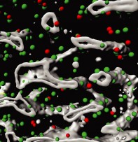
NK cells (red) attack normal leukemia cells (green). Leukemia stem cells (blue), on the other hand, suppress NKG2DL expression at their surface and thus escape destruction by the immune system. © Schürch/Lengerke, University and University Hospital of Basel

Leukemia stem cells protect themselves against the immune defense by suppressing a target molecule for natural killer (NK) cells. This protective mechanism can be tricked with drugs. The exciting finding has been made by colleagues of the Trumpp team from HI-STEM and DKFZ together with the laboratories of Claudia Lengerke (Basel) and Helmut Salih (Tübingen) and was now published in Nature. Together with our collaborators, we now want to develop new therapeutic approaches based on these results.

Update: July 26,2019: The article has been featured in Research Watch at Cancer Discovery: PARP1 Inhibition Overcomes Immune Escape of Leukemic Stem Cells from NK cells

Read more:

  • DKFZ Press Release
  • Read the Paper at Nature:
    Anna M. Paczulla, Kathrin Rothfelder, Simon Raffel, Martina Konantz, Julia Steinbacher, Hui Wang, Claudia Tandler, Marcelle Mbarga, Thorsten Schaefer, Mattia Falcone, Eva Nievergall, Daniela Dörfel, Pauline Hanns, Jakob R. Passweg, Christoph Lutz, Juerg Schwaller, Robert Zeiser, Bruce R. Blazar, Michael A. Caligiuri, Stephan Dirnhofer, Pontus Lundberg, Lothar Kanz, Leticia Quintanilla-Martinez, Alexander Steinle, Andreas Trumpp, Helmut R. Salih, Claudia Lengerke: Absence of NKG2D ligands defines leukaemia stem cells and mediates their immune evasion.
    Nature 2019, DOI: 10.1038/s41586-019-1410-1
  • Research Watch at Cancer Discovery: PARP1 Inhibition Overcomes Immune Escape of Leukemic Stem Cells from NK cells

On June 4th, as part of her visit of the DKFZ, her Honour Lieutenant Governor of Ontario Elizabeth Dowdeswel (@LGLizDowdeswell) visited the HI-STEM Lab. As part of her tour she showed great interest in our research on the role of stem cells in cancer.


https://twitter.com/LGLizDowdeswell

https://twitter.com/LGLizDowdeswell

Elizabeth Dowdeswell and Andreas Trumpp

Elizabeth Dowdeswell and Andreas Trumpp

Elizabeth Dowdeswell in the HI-STEM Lab

Elizabeth Dowdeswell in the HI-STEM Lab

Plot

The mammalian blood system is one of the best-researched hierarchical stem cell systems. It is also a main focus of the research in HI-STEM as it is a key for the understanding of the development of hematopoietic stem cells (HSCs) and the catastrophic deregulation of the system during e.g. development of leukemia.

An essential research tool in stem cell research is the ability to differentiate in detail the cell populations that make up the hematopoietic system. Today the method-of-choice is the separation of bone marrow cells via FACS (Fluorescence-Activated Cell Sorting). Here a population of cells is labelled with fluorochrome-conjugated antibodies against specific marker proteins, which are only expressed during certain stages of the hematopoietic development. However, separating the first stages of the system is extremely tricky as only single marker genes change between the stages and only certain combinations of fluorochromes can be applied due to overlaps in their fluorescence characteristics.

To improve and better standardize such approaches, HI-STEM researcher Marcus Eich and Steffen Schmitt (head of the DKFZ’s Core Facility Imaging and Cytometry), developed a new optimized multicolor immunofluorescence panel termed OMIP-059. The panel allows the simultaneous discrimination and quantification of all major populations of the mouse hematopoietic system from the earliest HSCs to the committed progenitor populations with high resolution using only one staining. In addition, it also integrates detection of the internal reporter gene GFP and the CD45.1 and CD45.2 alleles, which are frequently used for various applications.

All experimental details for the panel have now been published in the journal Cytometry: Part A so that the panel can be widely applied in HSC research.


Citation:

Eich, M., Trumpp, A., & Schmitt, S. (2019). OMIP-059: Identification of Mouse Hematopoietic Stem and Progenitor Cells with Simultaneous Detection of CD45.1/2 and Controllable Green Fluorescent Protein Expression by a Single Staining Panel. Cytometry A. doi: 10.1002/cyto.a.23845

Subcategories